")
Optical Tomography OCT

Optical Coherence Tomography is now an essential tool for a modern ophthalmology institute as it is the main method of diagnosis and continuous monitoring of all retinal diseases such as macular degeneration, diabetic retinopathy, central serous retinopathy, the early diagnosis of optic diseases nerve such as glaucoma, problems of the front part of the eye.
What is OCT
Optical coherence tomography (OCT) is the newest imaging method of the macula, optic nerve, cornea, optic papilla and anterior angle of the eye. It is a revolutionary method of “mapping” the anterior and posterior structures of the eye with detail corresponding to a histopathological preparation.
OCT Cornea
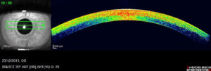 Optical Coherence Tomography (OCT) is the newest imaging method of the cornea of the eye. It is a revolutionary method of “mapping” the anterior structures of the eye with detail corresponding to a histopathological preparation.
Anterior Angle OCT
![]() Optical Coherence Tomography (OCT) is the newest imaging method of the anterior corner of the eye. It is a revolutionary method of “mapping” the anterior structures of the eye with detail corresponding to a histopathological preparation.
Optical Coherence Tomography (OCT) is the newest imaging method of the anterior corner of the eye. It is a revolutionary method of “mapping” the anterior structures of the eye with detail corresponding to a histopathological preparation.
OCT of the Optic Nerve Nipple
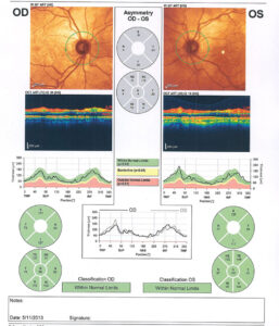 Optical Coherence Tomography (OCT) is the newest imaging method of the Papilla of the optic nerve of the eye. It is a revolutionary method of “mapping” the posterior structures of the eye with detail corresponding to a histopathological preparation.
Macula & Retina OCT
 Optical Coherence Tomography (OCT) is the newest imaging method of the macula and retina (retinal vessels) of the eye. It is a revolutionary method of “mapping” the posterior structures of the eye with detail corresponding to a histopathological preparation.